42 eye diagram with labels and functions
Generate eye diagram - MATLAB eyediagram - MathWorks Description. eyediagram (x,n) generates an eye diagram for signal x, plotting n samples in each trace. The labels on the horizontal axis of the diagram range between -1/2 and 1/2. The function assumes that the first value of the signal and every n th value thereafter, occur at integer times. eyediagram (x,n,period) sets the labels on the ... eyediagram - lost-contact.mit.edu eyediagram (x,n) creates an eye diagram for the signal x, plotting n samples in each trace. n must be an integer greater than 1. The labels on the horizontal axis of the diagram range between -1/2 and 1/2. The function assumes that the first value of the signal, and every n th value thereafter, occur at integer times.
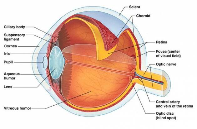
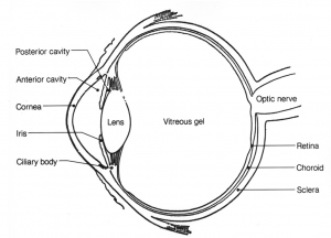
Human Eye: Structure of Human Eye (With Diagram) | Biology The human eye is a very sensitive and delicate organ suspended in the eye socket which protects it from injuries. It essentially consists of CORNEA, LENS & RETINA besides many other parts such as Iris, Pupil and aqueous humour, vituous humour etc. Each one has got a specific function. A section of the eye is as shown in Fig. 2.2. ADVERTISEMENTS:

Eye diagram with labels and functions
Microscope Parts and Functions With Labeled Diagram and ... Microscope Parts and Functions With Labeled Diagram and Functions How does a Compound Microscope Work?. Before exploring microscope parts and functions, you should probably understand that the compound light microscope is more complicated than just a microscope with more than one lens.. First, the purpose of a microscope is to magnify a small object or to magnify the fine details of a larger ... Eye Diagram With Labels and detailed description A brief description of the eye along with a well-labelled diagram is given below for reference. Well-Labelled Diagram of Eye The anterior chamber of the eye is the space between the cornea and the iris and is filled with a lubricating fluid, aqueous humour. The vascular layer of the eye, known as the choroid contains the connective tissue. en.wikipedia.org › wiki › DopamineDopamine - Wikipedia Dopamine is also synthesized in plants and most animals. In the brain, dopamine functions as a neurotransmitter—a chemical released by neurons (nerve cells) to send signals to other nerve cells. Neurotransmitters are synthesized in specific regions of the brain, but affect many regions systemically.
Eye diagram with labels and functions. Microscope Types (with labeled diagrams) and Functions Simple microscope labeled diagram Simple microscope functions It is used in industrial applications like: Watchmakers to assemble watches Cloth industry to count the number of threads or fibers in a cloth Jewelers to examine the finer parts of jewelry Miniature artists to examine and build their work Also used to inspect finer details on products Labelling the eye - Science Learning Hub In this interactive, you can label parts of the human eye. Use your mouse or finger to hover over a box to highlight the part to be named. Drag and drop the text labels onto the boxes next to the eye diagram If you want to redo an answer, click on the box and the answer will go back to the top so you can move it to another box. Structure and Functions of Human Eye with labelled Diagram Structure and Functions of Human Eye with labelled Diagram Biology Biology Article Structure Of Eye Structure of the Eye The eye is one of the sensory organs of the body. In this article, we shall explore the anatomy of the eye The structure of the eye is an important topic to understand as it one of the important sensory organs in the human body. Eye muscles and their functions - All About Vision The inferior oblique eye muscle originates from the front of the orbital floor, close to the nose. Its main function is to extort the eye when looking straight ahead (rotate the 12 o'clock position of the vertical meridian of the cornea toward the ear). It also elevates and abducts the eye (moves the direction of gaze upward and outward).
plotly.com › python › parallel-categories-diagramParallel categories diagram in Python - Plotly Basic Parallel Categories Diagram with graph_objects¶ This example illustrates the hair color, eye color, and sex of a sample of 8 people. The dimension labels can be dragged horizontally to reorder the dimensions and the category rectangles can be dragged vertically to reorder the categories within a dimension. Cow's Eye Dissection - Eye diagram - Exploratorium A muscle that controls how much light enters the eye. It is suspended between the A cow's iris is brown. many colors, including brown, blue, green, and gray. A clear fluid that helps the cornea keep its rounded shape. The pupil is the dark circle in the center of your iris. It's a hole that Your pupil is round. Eye Anatomy Diagram - EnchantedLearning.com Aqueous humor - the clear, watery fluid inside the eye. It provides nutrients to the eye. Astigmatism - a condition in which the lens is warped, causing images not to focus properly on the retina. Binocular vision - the coordinated use of two eyes which gives the ability to see the world in three dimensions - 3D. Cones - cells the in the retina that sense color. › graphs › process-flowCreate a Briliant Process Flow Diagram with Canva Process flow diagrams illustrate how a large complex process is broken down into smaller functions and how these fit together. As visual tools, they can help your team or organization see the bigger picture as well as where they fit into its entirety. Create a process flow any time you want to illustrate the stages of a process.
Draw a labeled diagram of human eye. Write the functions ... Cornea of the eye is the first sight where convergence of light rays takes place. Iris is that part of the eye which controls the amount of light entering the eye through the pupil. Pupil is a type of small hole through which light enters the eye. The eye lens is a convex lens just behind the pupilwhich converges the light rays towards the ratina. lisbdnet.com › diagram-of-how-blood-flows-throughdiagram of how blood flows through the heart – Lisbdnet.com Nov 28, 2021 · Essentially it is a muscle which functions as a really powerful pump. The heart takes in blood low in oxygen from the body. It pumps it through the right side of the heart and on to the lungs. In the lungs the blood passes through very small blood vessels and absorbs oxygen. What is the cardiac cycle GCSE? Eye Anatomy: 16 Parts of the Eye & Their Functions The following are parts of the human eyes and their functions: 1. Conjunctiva The conjunctiva is the membrane covering the sclera (white portion of your eye). The conjunctiva also covers the interior of your eyelids. Conjunctivitis, often known as pink eye, occurs when this thin membrane becomes inflamed or swollen. PDF Parts of the Eye - National Eye Institute Parts of the Eye . To understand eye problems, it helps to know the different parts that make up the eye and the functions of these parts. Here are descriptions of some of the main parts of the eye: Cornea: The cornea is the clear outer part of the eye's focusing system ... Eye Diagram Handout Author:
The eye, rods and cones - Biology Notes for IGCSE 2014 #88 Structure and function of the eye, rods and cones You need to be able to label parts of the eye on diagrams. The eyebrow stops sweat running down into the eye. Eyelashes help to stop dust blowing on to the eye. Eyelids can close automatically (blinking is a reflex) to prevent dust and other particles getting ton to the surface of the cornea.
Parts of Stereo Microscope (Dissecting microscope ... Apart from magnification, some eyepieces come with the label "WF" which defines that the eyepiece provides a wide field of view. This means that while viewing the specimen, the user will see wider areas compared to the field of view perceived using other eyepieces. Some of the most common field numbers are 18 and 20mm.
› TechnicalDocumentLibrary › GrafikControl Unit Installation and Operation Guide Please Read between any Eye QS control unit and any other power supply, including another GRAFIK Eye QS control unit. Refer to the QS Link Power Draw Units specification submittal (Lutron P/N 369405) for more information concerning PDUs. 1234 12 ABC 123456LN Example: Emergency lighting interface (maximum 1) Note: The GRAFIK Eye QS control unit
Anatomy of the Eye Diagrams for Coloring/Labeling, with ... Description. This printable contains 13 clear and simple cross sectional diagrams of the human eye. They photocopy well and are great for use as a labeling and coloring exercise for your students. The core eye anatomy diagram, designed as the labeling exercise, has a fully colored and labeled reference chart to go with it.
Parts of the Eye & Their Function | Robertson Optical and ... Eye Parts Description and Functions; Cornea: The cornea is the outer covering of the eye. This dome-shaped layer protects your eye from elements that could cause damage to the inner parts of the eye. There are several layers of the cornea, creating a tough layer that provides additional protection. These layers regenerate very quickly, helping ...
› consumers › consumer-updatesConsumer Updates | FDA Science-based health and safety information you can trust.
Human Eye Diagram, How The Eye Work -15 Amazing Facts of Eye First, light rays enter the eye through the cornea, the clear front "window" of the eye. The dome shaped cornea bends light to help the eye focus. From the cornea, the light passes through an opening called the pupil. The amount of light passing through is controlled by the iris, or the colored part of your eye.
Eye Diagram - Labelled Diagram of Human Eye, Explanation ... The basic functions of Rods and Cones are conscious light perception, color differentiation and depth perception. The human eye is capable of distinguishing between about 10 million colors, and it can also detect a single photo. The human eye is a part of the sensory nervous system. Labeled Diagram of Human Eye
Eye Diagram: Label Quiz - PurposeGames.com Layers of the Atmosphere 5p Image Quiz. 20 Major Rivers of the World 20p Image Quiz. Movie Quotes 6p Matching Game. Characteristics of Functions 10p Matching Game. Solar System Symbols 12p Image Quiz. Match the fractions, percentage and decimals 8p Matching Game.
PDF Eye Anatomy Handout - National Eye Institute of light entering the eye. Lens: The lens is a clear part of the eye behind the iris that helps to focus light, or an image, on the retina. Macula: The macula is the small, sensitive area of the retina that gives central vision. It is located in the center of the retina. Optic nerve: The optic nerve is the largest sensory nerve of the eye.
Eye anatomy and function - AboutKidsHealth Eye anatomy and function By SickKids staff. Listen Focus. download_for_offline Download PDF print_for_offline Print. An overview of how the many parts of the eye work together to produce clear vision. Key points. Visual acuity (VA) is defined as the clarity of the image seen by the eye. Visual acuity is measured using an eye chart at a distance ...
Eye Parts Labeling and Functions Flashcards - Quizlet layer of cells on the back of the eye cornea function helps protect the eye, and bends light to make an image appear on the retina through the lens iris function controls how much light enters the eye lens function makes an image on the eye's retina and can focus on objects that are close and far away by changing shape optic nerve function
ellenjmchenry.com › uploads › 2016CUT-AND-ASSEMBLE PAPER EYE MODEL of how the eye works, try one of these videos on YouTube. (Just use YouTube’s search feature with these key words.) “Anatomy and Function of the Eye: posted by Raphael Fernandez (2 minutes) “Human Eye” posted by Smart Learning for All (cartoon, 10 minutes) “A Journey Through the Human Eye” posted by Bausch and Lomb (2.5 minutes)
Label the Eye - The Biology Corner Label the Eye Shannan Muskopf December 30, 2019 This worksheet shows an image of the eye with structures numbered. Students practice labeling the eye or teachers can print this to use as an assessment. There are two versions on the google doc and pdf file, one where the word bank is included and another with no word bank for differentiation.
Structure And Function Of The Eye - Vision - MCAT Content The human eye is an organ that reacts with light and allows light perception, color vision, and depth perception. The photoreceptive cells of the eye, where transduction of light to nervous impulses occurs, are located in the retina (shown in Figure 1) on the inner surface of the back of the eye.But light does not impinge on the retina unaltered.
Microscope, Microscope Parts, Labeled Diagram, and Functions Microscope, Microscope Parts, Labeled Diagram, and Functions What is Microscope? A microscope is a laboratory instrument used to examine objects that are too small to be seen by the naked eye. It is derived from Ancient Greek words and composed of mikrós, "small" and skopeîn,"to look" or "see".
Generate eye diagram - MATLAB eyediagram eyediagram (x,n,period) sets the labels on the horizontal axis to the range between - period /2 to period /2. eyediagram (x,n,period,offset) specifies the offset for the eye diagram. The function assumes that the ( offset + 1)th value of the signal and every n th value thereafter, occur at times that are integer multiples of period.


Post a Comment for "42 eye diagram with labels and functions"